Sorted by what the tissue was doing
Ferdinand von Hebra's reorganisation of skin disease around pathological anatomy discarded centuries of visual cataloguing and built dermatology on the same intellectual foundation as the rest of medicine.
A catalogue with no principle
Before Hebra, the classification of skin diseases was essentially a problem of naming surfaces. Conditions were sorted by what a lesion looked like to the eye — its colour, its form, whether it was raised or flat, wet or dry. The Viennese physician Joseph Jakob Plenck had issued a taxonomy in 1776 that sorted eruptions into fourteen morphological classes; Alibert in Paris drew his celebrated nosological tree in the early nineteenth century, arranging diseases into families by their visible relationships. Both systems were rational within their own logic, and neither could explain why two conditions that looked nothing alike might arise from the same tissue process, or why two that looked identical might be entirely unrelated. The list that came before was a guide to appearances, not to causes, and it told a student nothing about what the skin was actually doing beneath the surface.

The problem was not merely intellectual. A student trained on morphological catalogues encountered a lesion and matched it to a picture. That match was as far as the reasoning went. There was no mechanism to incorporate new findings, no way to discard a category that turned out to contain unrelated things, and no connection to the pathological anatomy that was, by the 1840s, transforming the rest of Viennese medicine. The Allgemeines Krankenhaus — the great general hospital on the north side of the city — had become by mid-century one of the most powerful clinical research environments in Europe, and the anatomical school of Carl von Rokitansky was its engine. Hebra trained in that environment and drew the obvious conclusion: skin disease should be accounted for by the same framework that was reorganising internal medicine.

What Hebra changed, and how
Ferdinand von Hebra was appointed to run the skin ward at the Allgemeines Krankenhaus in 1845, and he set about reorganising the classification of skin disease around pathological anatomy — meaning the changes that could be observed in tissue, rather than the changes visible to the naked eye on the surface. A condition was not a type of rash. It was a type of tissue event: inflammatory, hypertrophic, atrophic, neoplastic. Two conditions that produced similar surfaces might belong to entirely different categories once you asked what the tissue was doing. Conversely, conditions that looked unlike one another might fall into the same group because the underlying process was the same kind of thing.
Chronology
- 1776Plenck issues morphological taxonomy, fourteen classes
- 1845Hebra appointed to the skin ward at the Allgemeines Krankenhaus
- 1856First parts of the Atlas der Hautkrankheiten issued
- 1870sKaposi integrates histological microscopy into the framework
This reorganisation had practical consequences that extended well beyond taxonomy. It meant that dermatology shared a language with the rest of medicine. A skin disease characterised by chronic inflammation occupied the same conceptual space as an inflammatory disease of any other organ. The discipline became, in principle, teachable by the same method — observation of process rather than memorisation of appearances — and the ward at the Allgemeines Krankenhaus became the site where that method was demonstrated daily. Students and visiting physicians came from across Europe to see it in action. The ward was large, the patient population was enormous, and Hebra used both to show that the same condition could be identified by its tissue logic across wildly varying presentations.
The classification and its instruments
Publishing the new system required a format that could carry both the argument and its evidence. The Atlas der Hautkrankheiten — issued in parts from 1856 onward — was the vehicle. Its plates were not decorative illustrations of surface appearances; they were records organised according to the new categories, showing conditions grouped by their pathological character. Anton Elfinger, the draughtsman who produced the original drawings, worked directly from patients on the wards. His training as both a medical artist and a caricaturist gave him an unusual capacity to render structure rather than merely surface, and the plates that resulted carry a precision that serves the taxonomic argument. The atlas came out in pieces over nearly two decades, and the classification it embodied was revised and extended as new evidence came in — an ongoing demonstration that the system was open to revision in a way the old morphological catalogues were not.

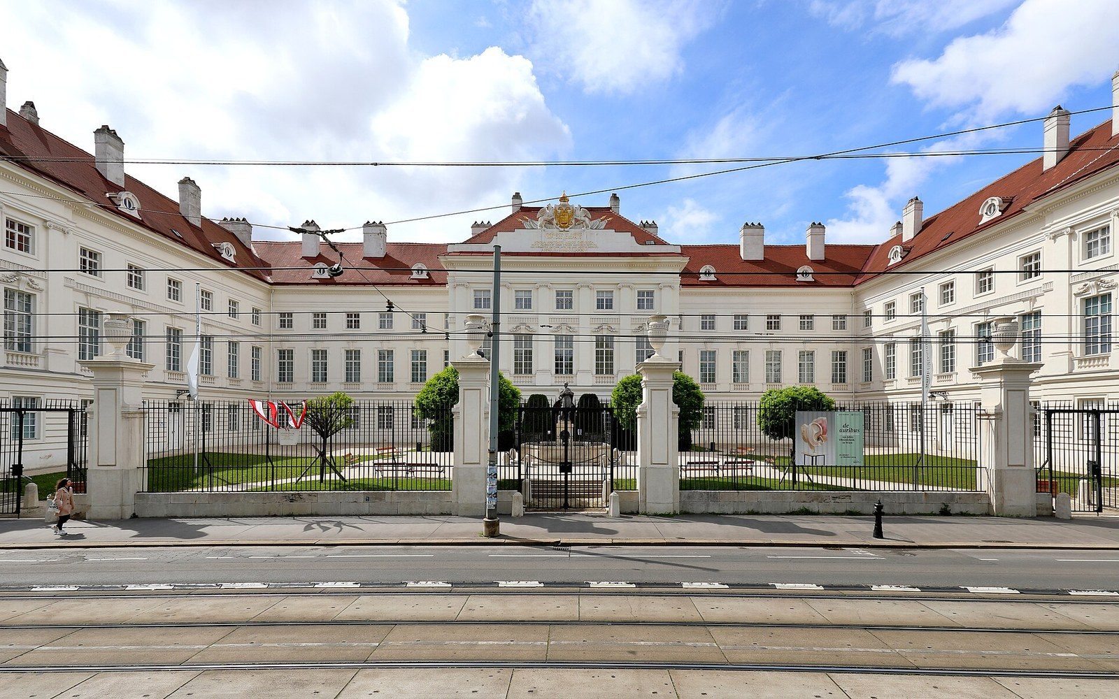
Where print met its limits, wax did the work. The moulage — a cast taken from a patient, filled with coloured wax, and painted to match the living skin — could record texture, elevation and three-dimensional structure in ways that even the finest lithographic plate could not reproduce. The collection built up in Vienna in the second half of the nineteenth century served the same teaching purpose as the atlas: to let a student encounter a condition without the patient being in the room. But crucially, the moulages were organised and labelled according to Hebra's categories, not according to the old visual typologies. A student consulting the collection was directed to ask not what the condition looked like but what the tissue was doing. The cabinet at the Josephinum holds part of this inheritance — objects whose arrangement still reflects the intellectual structure Hebra imposed.
How the system worked
- Old basis: visual morphology — colour, form, wet/dry, raised/flat
- New basis: tissue process — inflammatory, hypertrophic, atrophic, neoplastic
- Consequence: shared language with the rest of pathological anatomy
- Instruments: the atlas (print) and the moulage collection (wax)
Moriz Kaposi, who succeeded Hebra and married his daughter, extended the system in print. His own publications built on the pathological-anatomical framework while deepening the histological detail that microscopy was beginning to make available. By the 1870s, the school Hebra had founded was producing work that integrated gross pathological observation with microscopic tissue analysis, and the classification had absorbed that expansion without breaking. It was robust enough to take new evidence because it had been built on process rather than appearance.
Why it mattered beyond Vienna
The significance of what Hebra did extends past the internal history of dermatology. He demonstrated that a discipline organised around surface description could be reconstructed around mechanism, and that the reconstruction produced a more powerful clinical tool. The framework he imposed made it possible to teach systematically, to publish cumulative knowledge, and to connect findings across institutions that had never seen the same patient. It also made possible the kind of comparative teaching across a large and varied clinical population that the Allgemeines Krankenhaus uniquely afforded. The atlas and the moulage collection were not illustrations of the new system; they were its instruments, the means by which a conceptual reorganisation became a transmissible discipline.