Why photography could not do it yet
The lithographic atlas held on for two decades after the camera arrived — because early photography had nothing to offer a physician who needed colour.
The Medium and Its Limits
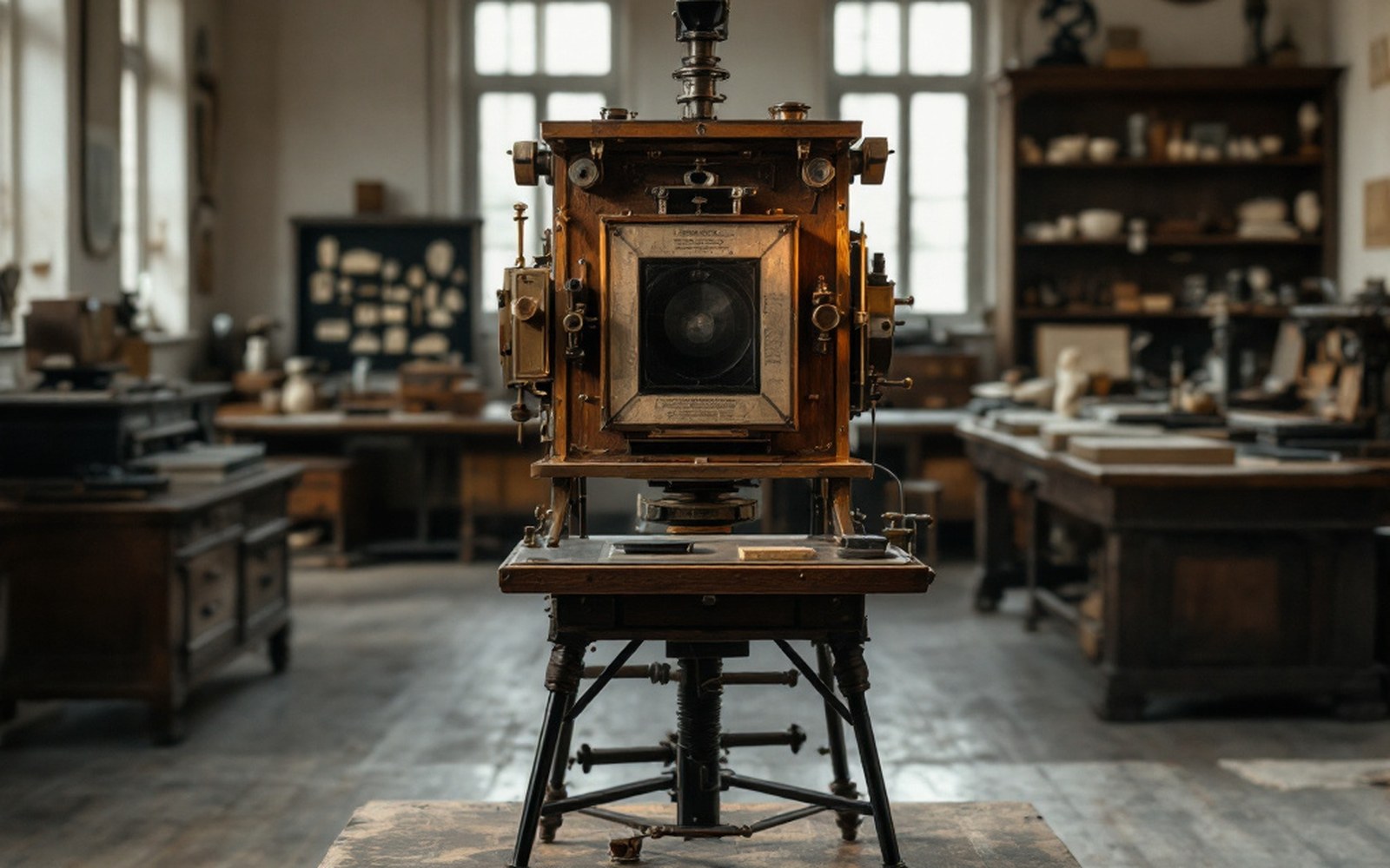
Photography reached Vienna in the early 1840s, within a year or two of Daguerre's announcement in Paris, and the city took to it quickly. Josef Petzval designed a faster portrait lens there in 1840, substantially reducing exposure times. Yet Ferdinand von Hebra, assembling the plates for his great atlas of skin diseases through the 1850s and 1860s, never considered replacing Anton Elfinger's drawn stones with photographic reproductions. The reason was not conservatism. It was physics.
The earliest processes — daguerreotype, then calotype, then the wet collodion plates that dominated professional photography into the 1870s — recorded the world in silver. Light struck a sensitized surface and produced tones of grey, and that was all. A physician trying to understand the difference between two superficially similar presentations that were pathologically distinct needed colour, or at least reliably reproducible colour. The lithographic plate, hand-coloured from a master copy, could give him that. The photograph could not.

There was a subtler problem beyond the absence of colour. Early photographic emulsions — all of them blue-sensitive only in this period — responded unevenly to the visible spectrum. They were acutely sensitive to blue and violet light while remaining almost blind to green and red. The consequence for a medical image was serious: tones that were in clinical fact quite different could collapse into the same grey. A patient's complexion, the precise boundary between inflamed and unaffected tissue, the depth of a pigmented area — all of these required the eye and hand of a trained draughtsman to interpret and transcribe faithfully. Elfinger, who was also a working caricaturist of real skill, understood how to read a surface and translate it into a mark that carried diagnostic weight. No lens could make that judgment.
The Coloured Plate as Instrument
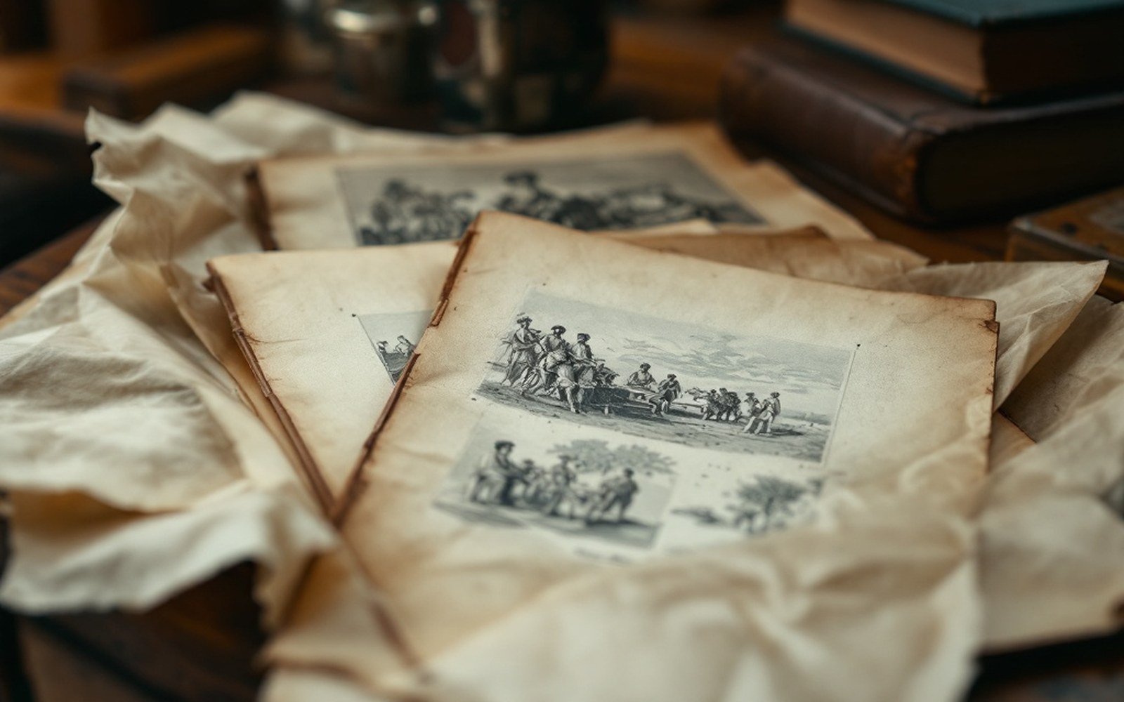
Hebra's atlas, issued in parts by the imperial publisher Wilhelm Braumüller across more than two decades beginning in 1856, reached subscribers as loose gatherings — unbound fascicles to be assembled later into complete volumes. Each plate arrived already coloured. That colouring was applied by hand after printing, worker by worker, each following the same master proof so that the image a subscriber in Prague received matched the one received in London. The system was labour-intensive and therefore expensive, but it was the only way to deliver a standardised, repeatable colour record at any scale. Photography, in the same decades, offered no equivalent.
Chronology
- 1839Daguerre announces photography in Paris
- 1840Josef Petzval designs the faster portrait lens in Vienna
- 1856Hebra's atlas begins publication in fascicle form, Wilhelm Braumüller, Vienna
- 1870sWet collodion plates still dominant in professional photography
- Late 1880s–1890sPhotomechanical halftone and photogravure processes reach practical use
The Allgemeines Krankenhaus in Vienna gave Hebra access to an extraordinary number and variety of patients, which is what made the atlas clinically authoritative. But access to patients does not produce a teaching image on its own. The translation from the ward to the printed plate required lithography — drawing directly onto prepared limestone and printing from it — precisely because lithography is a tonal medium capable of fine gradation, amenable to colour, and reproducible in editions large enough to reach a European medical audience. No photographic process of the 1850s or 1860s could print in colour at all, let alone in editions.
The Moulage as the Other Answer
Where even the coloured plate fell short — where surface texture and three-dimensional form mattered — the wax moulage (a cast taken from the patient's body, then painted) offered something photography could not attempt and print could only approximate. The collection at the Josephinum in Vienna preserves examples of this complementary technology: objects that are simultaneously scientific record and craft object, capturing what Hebra's atlas communicated across two dimensions. Photography would eventually threaten the moulage's role too, once colour processes matured and resolution improved. But throughout the period in which the atlas was being compiled and sold, the camera remained the wrong tool for the job, and working physicians knew it.
Photomechanical reproduction — the halftone screen and photogravure arrived in useful form only in the last decades of the nineteenth century, with colour processes such as autochrome following in the early twentieth. By then Hebra was dead, his atlas complete, and his successor Moriz Kaposi was working in a different technological moment. The atlas survived as a monument to the period when the draughtsman's hand was the only instrument precise enough to matter.